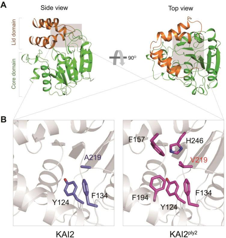
Fig. 6.
Three-dimensional X-ray crystal structure of KAI2 and KAI2ply2. (A) Left, a side view of KAI2. The ribbon representation of the overall structure of KAI2 is shown. Brown and green represent the lid and core domains, respectively. Right, a top view of KAI2: a 90° rotation of KAI2 relative to the side view is shown. (B) Magnified views of the areas in (A) shaded grey, showing residues forming hydrophobic interactions with A219 of KAI2 (left, indicated in blue) and V219 of KAI2ply2 (right, indicated in magenta).

