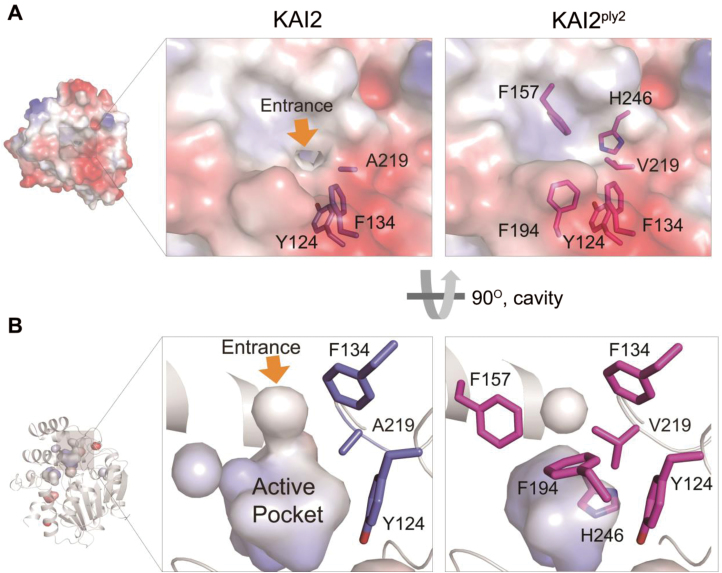
Fig. 7.
Structural comparison of KAI2 and KAI2ply2. (A) Surface views of KAI2 and KAI2ply2. KAI2 exhibits a small opening at the top, forming an entrance hole into the ligand-binding pocket (yellow arrow). Note that the entrance is closed in the KAI2ply2 protein. (B) Close-up view of the cavity on the active, putative ligand-binding pocket. Note that KAI2 shows an open-gate conformation with continuous space, whereas KAI2ply2 shows a closed gate with discontinuous space.

