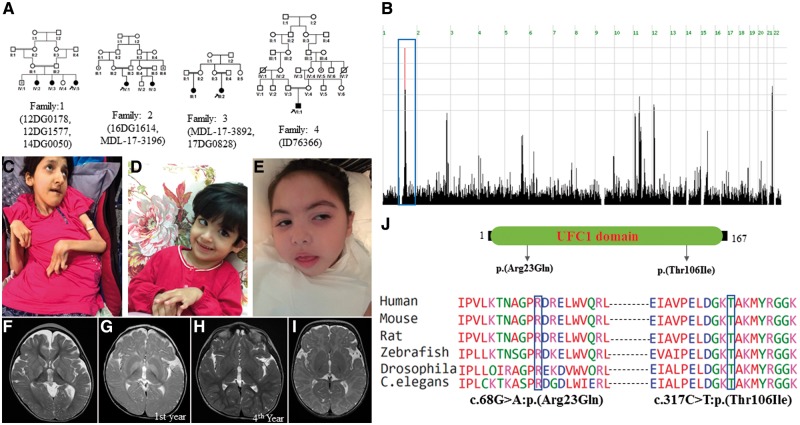
Figure 2.
UFC1-related clinical phenotype. (A) Pedigrees of the families with UFC1 mutation. (B) Genome-wide homozygosity plot highlighting a single critical locus from Families 1–3, who harbour the p.(Thr106Ile) mutation. Blue box indicates haplotype (Chr1:158957700-162094700) identical by descent. (C) Clinical images showing multiple joint contractures and emaciation (Patient 17DG0828). (D and E) Clinical images of younger patients suggest a progressive nature of emaciation (Patients 14DG0050 and 16DG1614, respectively). (F) Brain MRI of Patient 16DG1614 showing bilateral hyperintense signals in the basal ganglia. (G and H) Brain MRI of Patient MDL-17-3196 showing bilateral hyperintense signals in the basal ganglia in the first year that resolved in a repeated brain MRI in the fourth year of age. (I) Brain MRI of Patient ID76366 showing markedly delayed myelination. (J) Schematic diagram of UFC1 protein showing UFC1 domain and mutation site. Mutations Arg23Gln and Thr106Ile are highly conserved from humans to C. elegans.

