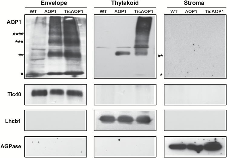
Fig. 3.
Localization of AQP1 in the thylakoid and envelope membranes. Envelope, thylakoid, and stroma fractions were isolated from wild-type (WT), AQP1, and TicAQP1 leaves, and separated by SDS-PAGE. Samples of 2, 20, and 30 μg of protein from the envelope, thylakoid, and stroma, respectively, were loaded per well. Representative western blots performed with antibodies to AQP1, the inner-membrane Tic40 protein, the thylakoid membrane-specific LHC chlorophyll a/b binding protein 1 (Lhcb1), and the stroma-specific ADP-glucose pyrophosphorylase (AGPase) are shown. Asterisks indicate the positions of monomer (*), dimer (**), trimer (***), and tetramer (****) AQP1.

