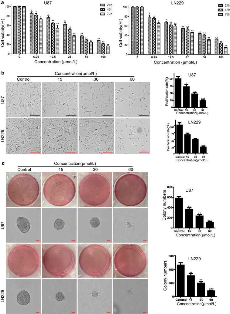
Fig. 2.
Compound-1H reduces cell proliferation and viability of human glioblastoma cells. a U87 and LN229 cells were treated with the different concentrations of compound-1H for 24, 48 and 72 h. Cell viability was measured with the MTT assay. b Cell morphology of U87 and LN229 glioblastoma cells was captured with microscope after treating with vehicle (0.05% DMSO) or the indicated concentrations of compound-1H for 48 h. Scale bar, 100 μm. The histogram showed the quantification of cell proliferation rate. c The soft agar assay was employed to detect colony formation in vitro after treating with the indicated concentrations of compound-1H for 14 days. The colonies were visualized with the images and quantitated by histogram. Scale bar, 100 μm. All data were demonstrated as the mean ± SD of three independent experiments. *P < 0.05; **P < 0.01; ***P < 0.001 versus vehicle

