
Figure 3. The preferential lipid boundaries increase CPR’s ability to transfer electrons to cyt c.
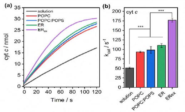
a) Kinetic traces of cyt c reduction as measured by UV-vis absorption spectroscopy. b) Calculated kcat for cyt c reduction by CPR in solution or in 4F-nanodiscs. Data are expressed as average ± standard deviation (n = 3). * p<0.05, ** p<0.01, *** p<0.001.