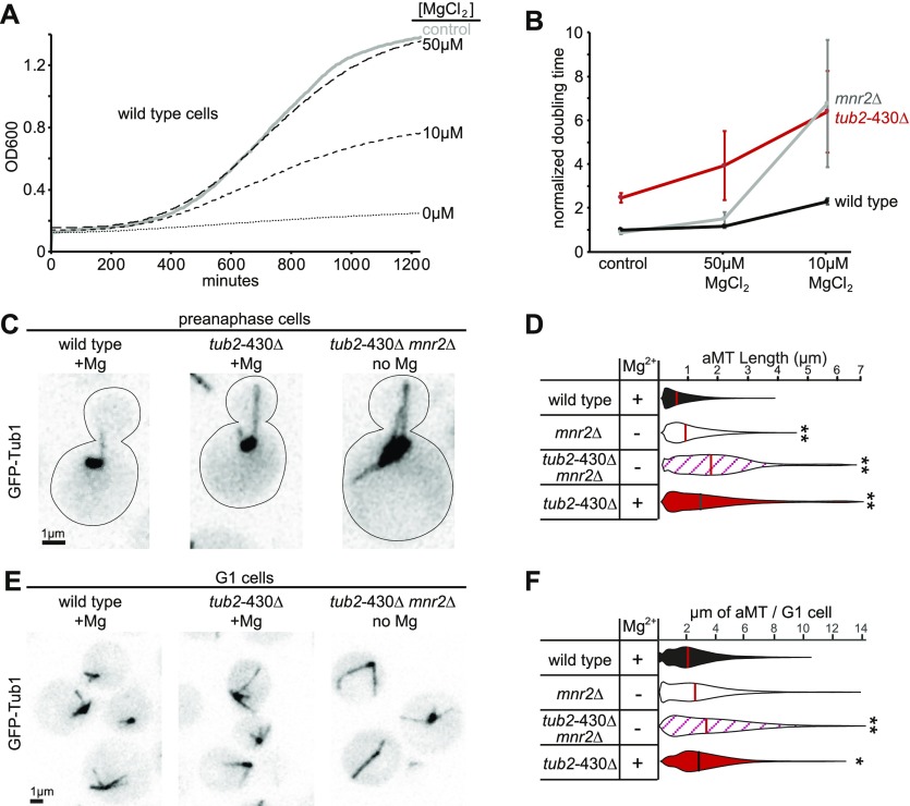
Figure 5. β-CTT regulates magnesium sensitivity in vivo.
(A) Representative growth curves of wild-type cells in different magnesium conditions. Control group was cultured in synthetic media with 4 mM MgSO4. Other groups were grown in synthetic media without MgSO4 but supplemented with MgCl2 at concentrations indicated. All cultures were grown at 30°C with agitation for 21 h, and OD600 was measured every 5 min. (B) Median doubling times of wild-type, mnr2Δ, and tub2-430Δ cells at indicated magnesium conditions, normalized to the doubling time of wild-type cells in 4 mM MgSO4. Each data point represents at least 11 replicates from four separate experiments. Error bars are ± 95% CI. (C) Representative images of cells in preanaphase expressing GFP-labeled microtubules, grown in synthetic media or without MgSO4. Scale bar = 1 μm. (D) Distribution of astral microtubule (aMT) lengths measured in preanaphase cells from an asynchronous culture. Lengths were measured every 4 to 5 s for 5 min. Cells were cultured in synthetic media with (+) or without (−) 4 mM MgSO4. Data pooled from two separate experiments, with at least seven cells and 396 total measurements for each group. **P « 0.001. Significance determined by the Mann–Whitney U test. Lines denote median. (E) Representative images of cells in G1 phase expressing GFP-labeled microtubules, grown in synthetic media or without MgSO4. Scale bar = 1 μm. (F) Distribution of aMT lengths measured in G1 from an asynchronous culture. Cells were cultured in synthetic media with (+) or without (−) 4 mM MgSO4. Data pooled from two separate experiments, with at least 60 cells for each group. *P = 0.01, **P « 0.001. Significance determined by the Mann–Whitney U test. Lines denote median.

