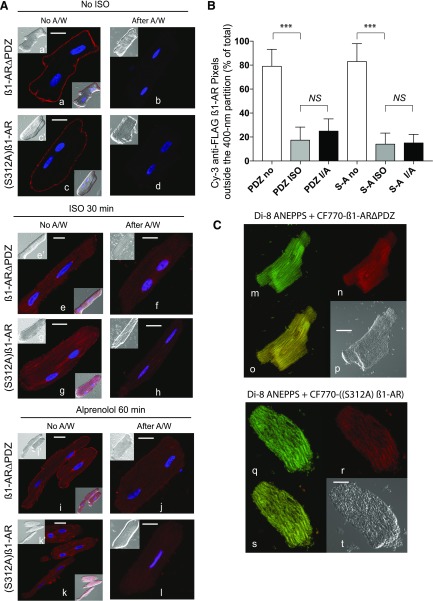
Fig. 3.
Compartmentalization of β1-AR∆PDZ and (S312A) β1-AR in ARVMs. (A) Confocal microscopy of FLAG β1-AR∆PDZ or FLAG (S312A) β1-AR in ARVMs was carried as previously described in Fig. 1A. Each scale bar in (A) represents 15 µm. (B) Pixel distribution of Cy3 β1-AR ∆PDZ (PDZ) or Cy3-[(S312A) β1-AR] (S-A) outside the 400-nm partition (mean ± S.E.) of ARVMs that were untreated and exposed to ISO or ISO/ALP by the microscopic partition procedure from 30 images/condition that were derived for three separate experiments is presented. Statistical comparisons were carried out by one-way ANOVA followed by Bonferroni’s test. NS, indicates nonsignificant differences between the column pairs or *, **, and *** to indicate P < 0.05, P < 0.01, and P < 0.001, respectively. (C) ARVMs expressing FLAG-β1-AR ∆PDZ or FLAG (S312A) β1-AR were processed as described in Fig. 2F. Slides were permeabilized and incubated with CF770-goat anti-rabbit IgG and visualized. β1-AR∆PDZ staining (pseudo red, image n) or (S312A) β1-AR (pseudo red, image r) and that of with Di-8 ANEPPS (images m and q) were superimposed (images o and s, respectively). Each scale bar in (A) and (B) represents 15 µm.

