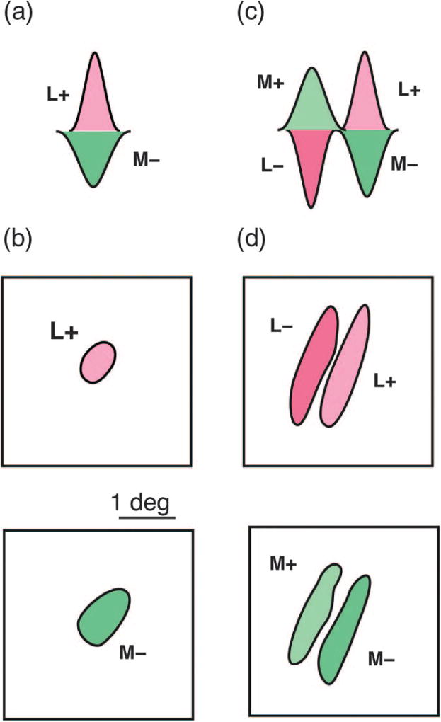
Fig. 5.
Spatial and chromatic opponent receptive fields. (a) Schematic spatial sensitivity profile of a single-opponent receptive field receiving excitatory input (L+) from long-wave sensitive cones, and inhibitory input (M−) from middle-wave sensitive cones. (b) The receptive field projection of a single-opponent field shows approximately overlapping and circularly symmetric cone-opponent inputs. (c) Schematic spatial sensitivity profile of a double-opponent receptive field. Here the receptive field comprises two opponent sub-regions which are spatially offset. (d) The receptive field projection of a double-opponent field shows complementary, orientation-selective sub-regions which produce both spatial and chromatic selectivity. Modified from [53].

