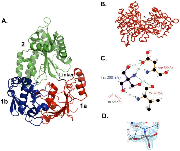
Figure 1.
X-ray crystal structure of E. coli MppA. (A) The open form of MppA is a pear-shaped, bilobed structure. Domain 1a is shown in red, 1b is in blue, and 2 is in green. The linker region is in black. (B) The Tris binding site. Contacts are made between a bound Tris molecule (black bonds) with amino acid residues in one copy of MppA (red, molecule A) in the asymmetric unit. (A,B were made using PyMol [43]). (C) The Tris buffer molecule interacts with the side chain of Asp439 and the backbone of Val437, near the sidechain of Trp436. (C was made with LigPlot within Procheck). (D) 2fo-fc Electron density map around bound Tris molecule contoured at 1.0 (made with NGL viewer [46,47]).

