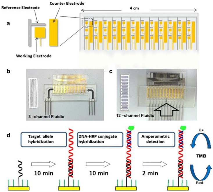
Figure 2.
Schematic of the electrochemical genosensor array: (a) the 36-electrode array and a zoomed view of the electrode arrangement; (b,c) electrode array after mounting within 3- and 12-channel fluidic cells; (d) steps involved in genosensor assay [45]. Copyright 2014. Reproduced with permission from Springer-Verlag.

