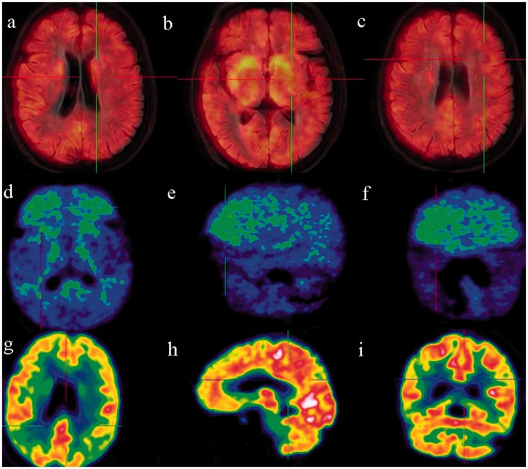
Figure 1.
A 70-year-old woman with a 3-year disease course. (a)–(c) DaT PET imaging revealed reduced uptake in the bilateral caudate nuclei and putamina. (d)–(f) PIB PET imaging revealed clear uptake in the frontal lobe and basal ganglia and a slight increase in the parietal, temporal, and occipital lobes. (g)–(i) FDG PET imaging revealed decreased metabolism in the frontal lobe and the junction between the parietal, occipital, and temporal lobes. FDG metabolism was normal in the occipital lobe, posterior cingulate cortex, basal ganglia, and cerebellum. DaT, dopamine transporter; PET, positron emission tomography; PIB, Pittsburgh compound B; FDG, fluorodeoxyglucose.

