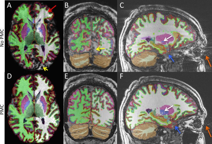
Fig 3.
MPRAGE axial (A and D), coronal, (B and E), and sagittal (C and F), images overlaid with segmentation results. Top and bottom rows are the No PMC and PMC conditions, respectively. The red arrow indicates ringing artifacts due to motion. The yellow arrows indicate areas of poor segmentation due to motion artifacts. The orange arrows indicate artifacts in the phase-encode direction due to eye movement. The blue arrows indicate areas of poor segmentation in the temporal lobe due to eye movement artifacts. The white arrows indicate both the under and over estimation that occurs in subcortical structures due to motion artifacts.

