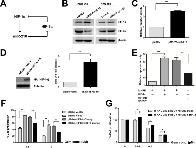
Fig 7. Suppression of miR-210 regulates HIF-1α activity via the function of HIF-3α.
(A) A schematic diagram shows the hypothetical role of miR-210 in regulating HIF-1α function. (B) Protein levels of HIF-1α and HIF-3α in the stably transfected CCA cells. (C) HRE (hypoxia responsive element)-reporter assay in KKU-213 cells induced by miR-210 expression vector. (D) The expression level of miR-210 in KKU-213 cells transiently overexpressed with HIF-1α vector. (E) The 293T cells were co-transfected with Luc-3xHRE, HIF-1α expression vector and miR-210 sponge as well as control vectors. At 72 h post-transfection, cells were lysed and luciferase activity was determined. Data were normalized with the luciferase activity of Renilla. (F) Cell proliferation assay in KKU-213 cells transiently transfected with HIF-1α and miR-210 sponge vector in responding to gemcitabine treatment. (G) Cell proliferation assay in the stable miR-210 overexpressing KKU-213 treated with si-HIF-3α and incubated with gemcitabine under pseudohypoxia for 72 h. The data present the mean ± SD of three-independent experiments. *P < 0.05. **P < 0.01. ***P < 0.001.

