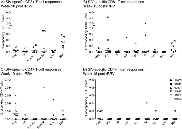
Fig 9. Intracellular cytokine staining analysis of vaccine-induced SIV-specific T-cell responses in rRRV-SIVcmv-nfl-inoculated macaques.
The magnitude and specificity of vaccine-elicited CD8+ (A) and CD4+ (B) T-cell responses were measured in PBMC by ICS using pools of peptides (15mers overlapping by 11 amino acids) spanning the appropriate SIVmac239 proteins. In the week 10 assay (A & C), Vpx and Vpr peptides were grouped in a single pool and so were the Rev and Tat peptides. Pol peptides were omitted in the week 10 assay. In the week 18 assay (B & D), 1–3 peptide pools corresponding to individual SIVmac239 proteins were used as stimuli. The percentages of responding CD8+ T-cells or CD4+ T-cells displayed were calculated by adding the frequencies of positive responses producing any combination of three immunological functions (IFN-γ, TNF-α, and CD107a). Lines represent medians and each symbol corresponds to one vaccinee.

