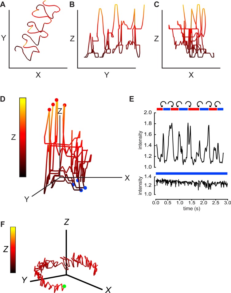
Fig 2. Digital holographic imaging also provides 4D description of sperm swimming trajectories and rolling behavior.
The path of the head of another free-swimming mouse sperm with a linear averaged trajectory was projected onto the XY-plane (A), the YZ-plane (B), and the XZ-plane (C). Equal scaling (14 μm) of all axes. Z-plane excursions are also color coded as indicated by the vertical bar in panel D below. The 2nd segment of S1 Movie is the XY projections of the 2.2s holographic record. (D) A 3D view of the swimming path, red balls indicate path locations where the cell rolls from “LCh” to “RCh” orientation, blue balls indicate locations where cell rolls “RCh” to “LCh”. (E) (upper panel) Red and blue bars indicate periods of “RCh” and “LCh” orientation for this cell. The orientation is indeterminate in the short white areas. Curved arrows indicate CW or CCW rolling around the long axis of the cell. Also shown is the oscillating intensity of light scattered from the head of the rolling sperm (relative to background from a cell-free area). In the lower panel, a different, non-rolling sperm maintains a LCh orientation and lacks periodic fluctuations in intensity of reflected light. (F) A 3D view of the circular path of this non-rolling cell. Green ball marks the starting point. Scale bars 14 μm. Z-plane excursions color coded as in D.

