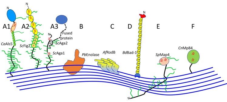
Figure 2.
Cartoon models of some fungal adhesins, illustrating different domain arrangements and cell wall associations. A cell wall is shown as blue lines, representing glucan polymers. For each cartoon, abbreviations for the genus and species are in italics: Ca, C. albicans; Sc, S. cerevisiae; Pb, Paracoccidioides braziliensis; Af, Aspergillus fumigatus; Blastomyces dermatitidis; Sp, Schizosaccharomyces pombe, Cn Cryptococcus neoformans. The name of each adhesin is given in Roman font. Hydrophobic domains are filled in yellow. Potential amyloid-forming β-aggregation core sequences are shown as red zigzags; O-linked glycosylations are short green lines, N-glycans are longer green lines. C represents Cys-rich sequences in ScFig2 (A2) and AfRodB (C), and CW the Cys/Trp-rich domains in Bad-1 (D). Adhesins labeled (A) are covalently attached to the wall through modified GPI anchors, and (F) may be as well. The other sub-figure indices (B through E) show other cell wall attachment modes and are described in the text.

