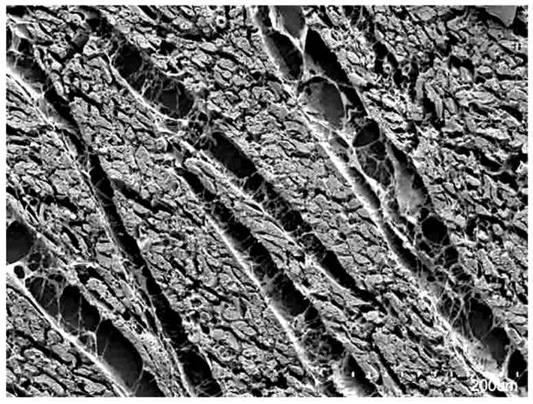
Figure 1.
The scanning electron micrograph, taken from a sub-volume of the ventricular wall, shows how the individual cardiomyocytes are aggregated together by the endomysial component of the fibrous matrix into units that are separated by perimysial clefts containing loose connective tissue. There is, however, no uniformity in the thickness of the aggregated units, which can be seen to be heterogeneous and interconnected branching entities.

