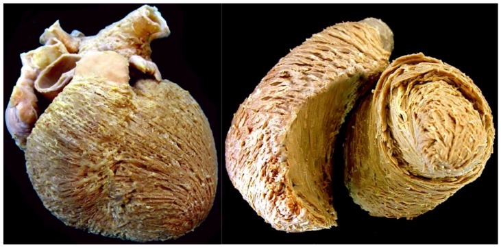
Figure 3.
The dissection shown in the (left hand) panel was made by removing or ‘peeling’ the epicardial covering of the ventricular cone. It shows the obvious “grain” produced by the aggregation of the cardiomyocytes into chains. Note that the superficial cardiomyocytes are shared between the ventricles, with no traceable division. The left ventricular chains show a marked angle relative to the ventricular long axis. The image shown in the (right hand) panel demonstrates that the cardiomyocytes in the left ventricular mid-wall are aggregated in circumferential fashion.

