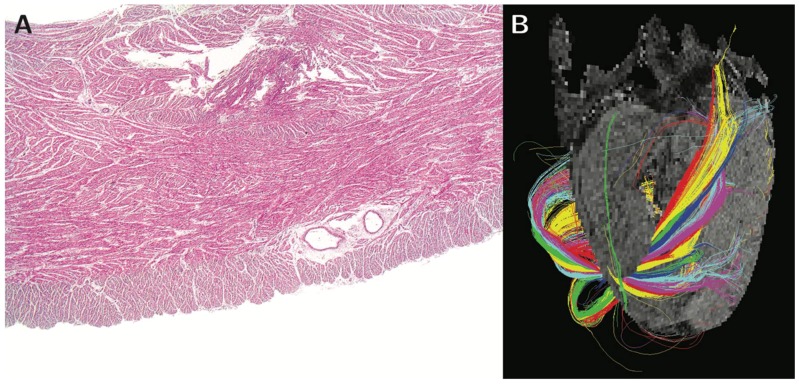
Figure 5.
Panel (A) shows a histological section of the right ventricular wall of the sheep heart stained with hemotoxylin and eosin, revealing the heterogeneous mural architecture. The helical angle of the chains of cardiomyocytes aggregated together within the right ventricular walls is shown in panel (B), which is a tractograph generated from different zones of the walls. The color coding of the tractography does not represent any anatomical or physiological property. It serves only as a visual aid enabling the reader to distinguish between the tracks, and to visualize the mean orientation of cardiomyocytes within them. Modified from Agger et al., 2015 [48].

