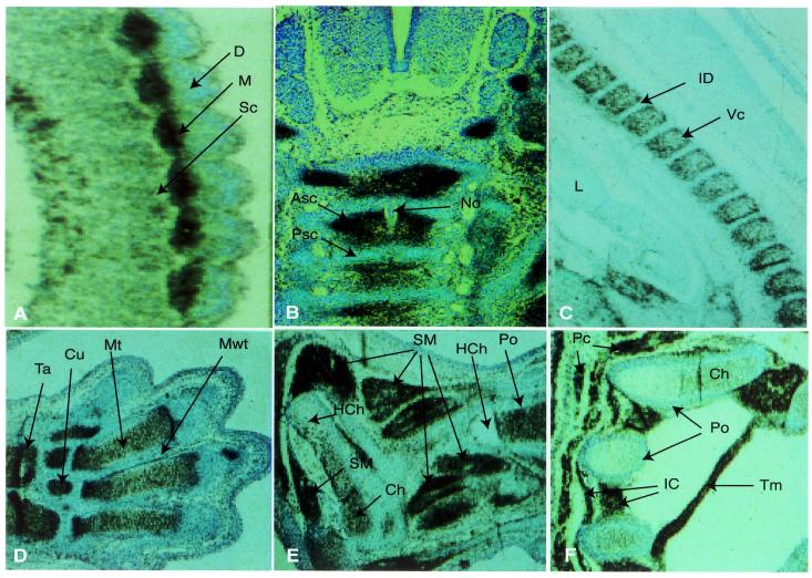
Figure 4.
Edr expression during musculo-skeletal development. Sagittal (A,C,D–F), or transverse (B) sections of E10.5 (A), E12.5 (B), E14.5 (C), E17.5 (D–F) mouse embryos were hybridised with antisense 35S-radiolabelled RNA probes derived from the Edr cDNA clone pGR165, subjected to autoradiography for 48 h and counter-stained with toluidine blue. (A–C) 100× magnification. (D–F) 40× magnification. Some folding of tissue is apparent in (D). ASc, anterior sclerotome; Ca, calcaneus; Ch, chondrocytes; Cu, cuneiforme; D, dermatome; HCh, hypertrophied chondrocytes; IC, intercostal muscle; ID, intervertebral disc; M, myotome; Mt, cartilage primordium of metatarsale; Mwt, mesodermal web tissue; No, notochord; Pc, cutaneous muscle of thorax and trunk (panniculus carnosus); Po, periosteum; PSc, posterior sclerotome; Ra, radius; Sc, sclerotome; SM, fetal limb skeletal muscle; Ta, talus; Tm, Transversus thoracis muscle; VC, vertebral cartilage.

