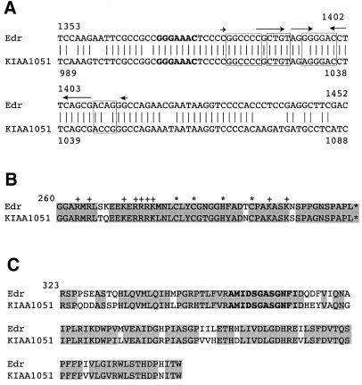
Figure 6.
Conservation of the –1 ribosomal frameshift signals and the aspartyl protease motif in human KIAA1051 gene. (A) Alignment of the nucleic acid sequences of Edr and KIAA1051 cDNAs. The heptameric –1 slippage sequence is denoted in bold. Conserved primary sequences for the potential pseudoknot and the simple stem–loop structures are shown in closed boxes, overlined (arrowed), respectively (KIAA1051 Genbank accession no. AB028974). (B) Comparison of the CCHC motif from Edr and KIAA1051. Conserved CCHC motifs are shown by asterisks. Basic residues outside CCHC motifs are marked with +. Identical residues are shaded. (C) Comparison of the putative aspartyl protease active sites from Edr and KIAA1051. Conserved residues for the aspartyl protease active sites are shown in bold. Identical residues are shaded.

