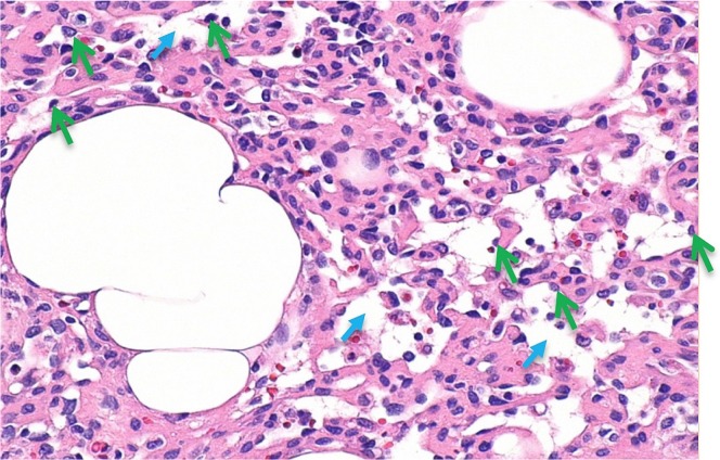
FIGURE 4.
High-magnification view of anastomosing channels with occasional red blood cells. The green arrows mark hobnail endothelial cells. The smaller blue arrows mark anastomosing channels. Hematoxylin and eosin stain. 20× original magnification. Horizontal dimension of the image is approximately 0.42 mm.

