
Figure 3.
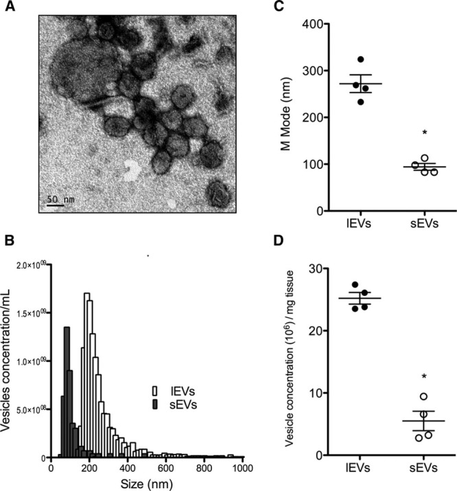
Intracardiac extracellular vesicles (EVs) in human myomectomy biopsies. A, Typical representative electronic microscopy of cardiac EVs from human biopsies indicating the presence of large (lEVs) and small EVs (sEVs). B, Representative size distribution analysis by tunable resistive pulse sensing (TRPS) of lEVs (in white) and sEVS (in grey). C, M-mode analysis of both lEVs and sEVs. D, EV concentration determined by TRPS. Data are mean±SEM. n=4 samples per fraction. *P<0.05 vs lEVs.