
Fig. 1.
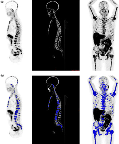
Quantification of tumoral bone burden in a prostate cancer patient with disseminated bone metastases: (a) bone volume measurement on whole-body computed tomography (CT) extrapolated on PET [sagittal view PET, right; sagittal view bone CT, center; maximum intensity projection (MIP), left]; (b) metabolic bone tumor volume measurement on PET with standardized uptake value>3, selected regions upon bone segmentation in blue: sagittal view PET on the right, sagittal view bone CT in the center, MIP on the left side.