Figure 2.

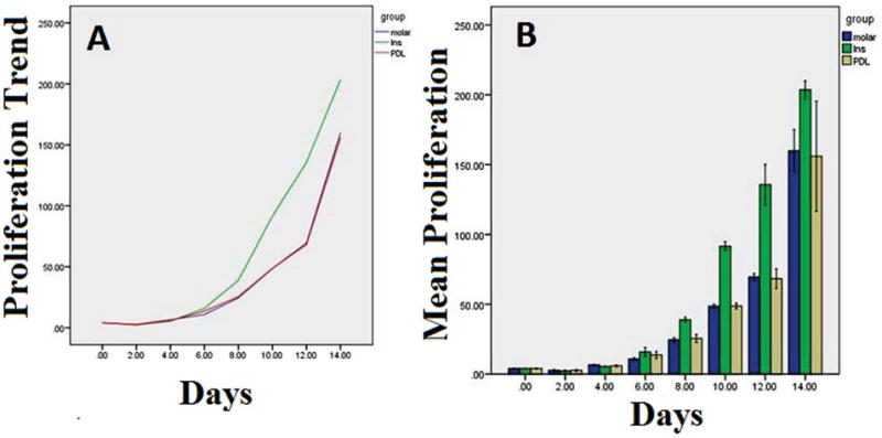
A: The trend of the proliferation rate of the ECs isolated from PDL, incisor (Ins) and molar teeth. B: The box plot of the means and standard deviations of the proliferation rate of the ECs isolated from PDL, incisor (Ins) and molar teeth.

A: The trend of the proliferation rate of the ECs isolated from PDL, incisor (Ins) and molar teeth. B: The box plot of the means and standard deviations of the proliferation rate of the ECs isolated from PDL, incisor (Ins) and molar teeth.