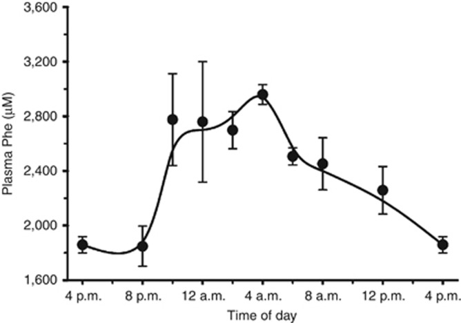
Figure 2.
Diurnal variation in plasma phenylalanine (Phe) levels of untreated female phenylketonuria (PKU) mice. The change in plasma Phe levels that occurred during the 24 h dark–light cycle was determined from analysis of composite data. Blood samples (40 μl) were obtained from six adult female PKU mice at the indicated times, with samples collected over a 9-week period to prevent stress from blood loss. Two samples were obtained from each animal at every time point. Plasma Phe results were combined and averaged to yield the composite 24 h data plot. The plot shows the fast rise of over 1,000 μM in plasma Phe that occurred when the mice woke at 6 PM, began feeding, and the slower fall beginning at the 6 AM start of the light cycle.

