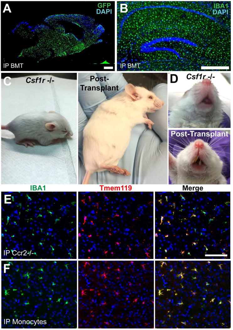
Figure 3. Peripheral bone marrow injection leads to widespread engraftment of donor-derived cells and results in partial rescue of the Csf1r−/− phenotype; both purified monocytes and Ccr2 −/− bone marrow cells engraft in the Csf1r −/− brain and express Tmem119.

A) Engrafted MLCs 1 month after intraperitoneal (IP) bone marrow injection into P1 host. Scale bar = 900μm. B) Hippocampal section of Csf1r−/− brain stained for IBA1 8 months after IP BMT. Scale bar = 400μm. C) Typical Csf1r−/− mouse showing abnormal head shape, small size (left), compared to 3 months after intraperitoneal bone marrow injection at P2 (right). D) Untransplanted Csf1r−/− mouse lacks teeth (top), while transplanted Csf1r−/− mouse shows tooth growth (bottom). E) Both Ccr2 Rfp/Rfp BM and F) purified BM monocytes engraft in the Csf1r −/− brain and express TMEM119 at T=21 days. Scale bar = 100μm. See also Figure S3.
