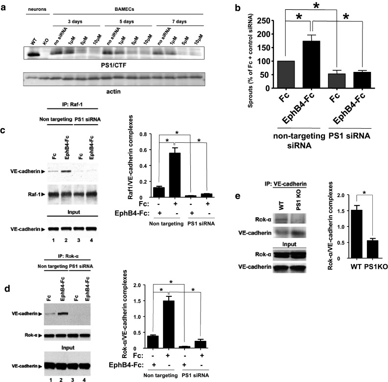
Fig. 6.

EphB4-Fc-induced sprouting and angiogenic complex formation depends on PS1. PS1 promotes angiogenic complexes in vivo. a Cells were nucleofected with various concentrations of PS1 siRNA or buffer alone. After the indicated days, cells were lysed in 1% SDS lysis buffer and immunoblotted with 33B10 mouse monoclonal antibodies against PS1/CTF and actin. Cortical neuronal extracts from PS1 WT and KO embryonic mice were used as a positive control for PS1. b Cells were nucleofected with 7.5 μM of bovine-specific PS1 siRNA or non-targeting siRNA. Cells were then used for the microcarrier bead assay in the presence of pre-clustered Fc or EphB4-Fc as described. The percentage of sprouts exceeding the diameter of the bead relative to control (Fc + non-targeting siRNA) was determined. Paired t test (n = 4, *p < 0.05, error bars = SEM). c Left: cells were nucleofected with PS1 siRNA or control non-targeting sequences as described in b and treated with 2 μg/ml Fc or EphB4-Fc as indicated. Cell extracts were IPed with anti-Raf1 antibody and IPs were probed on WB for VE-cadherin (upper panel) or Raf-1 (middle panel). Input panel shows expression of VE-cadherin. Right: quantification of results. Paired t test, n = 3, *p < 0.05, error bars = SEM. d Left: cell extracts from (c) were IPed with anti-Rok-α antbody and IPs were probed on WB for VE-cadherin (upper panel) or Rok-α (middle panel). Input panel shows expression of VE-cadherin. Right: quantification of results. Paired t test, n = 3, *p < 0.05, error bars = SEM. e Left: brain extracts from WT or PS1 KO mouse embryos were IPed with anti-VE-cadherin antibodies and IPs were probed on WB for Rok-α (first panel) or VE-cadherin (second panel). Input shows Rok-α and VE-cadherin expression. Right: quantification of results. Paired t test, n = 3, *p < 0.05, error bars = SEM
