
Figure 1. The RSK2-mediated inflammation pathway.
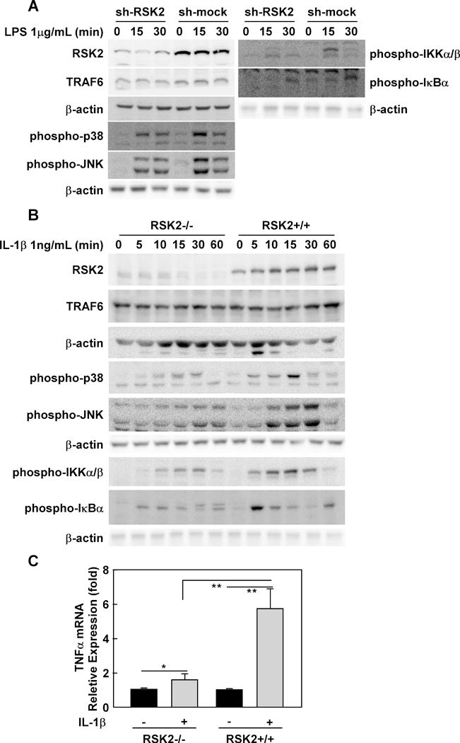
(A) RSK2 knockdown suppresses LPS-induced inflammation signaling. RAW cells were infected with an sh-mock or sh-RSK2 viral vector and each stable cell line was established. RSK2 knockdown was confirmed by Western blot (panel 7 from top). Sh-RSK2 and sh-mock cells were cultured and stimulated with LPS (1 μg/mL). Phosphorylated Ikkα/β, IKBα, JNKs, p38 and total TRAF6 proteins were visualized by Western blotting. (B) RSK2 deficiency blocks IL-1β–induced inflammation signaling. RSK2+/+ and RSK2−/− murine embryonic fibroblasts (MEFs) were cultured to 90–95% confluence, and subsequently starved in 0.1% FBS-DMEM for 24 h. The cells were stimulated with IL-1β (1 ng/mL) and harvested at the indicated time point. Western blotting was conducted using whole cell lysates from RSK2+/+ and RSK2−/− MEFs as indicated. The levels of phosphorylated Ikkα/β, IKBα, JNKs, p38 and total RSK2 proteins were visualized by Western blot. Each assay was performed 3 times and representative blots of similar results are shown. (C) RSK2 deficiency inhibits the induction of TNFα expression by IL-1β. Graph data are shown as means ± S.D. of values obtained from triplicate experiments and significant differences were evaluated using the Student’s t- test (*, p < 0.05; **, p < 0.01).