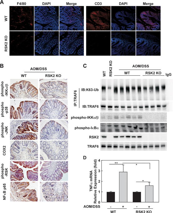
Figure 7. Less inflammation signaling is observed in RSK2 KO mice compared with wild type controls.

(A) F4/80 and CD3 are down-regulated in RSK2 KO mice. RSK2 KO and wild type control colon tissues were fixed and subjected to immunofluorescence analysis. Sections were stained as described in Methods using anti-F4/80 and anti-CD3 followed by Alexa568-conjugated goat anti-mouse IgG2A. Nuclei were counterstained with DAPI. Representative images from each group (n = 5) are shown. (B) RSK2 deficiency reduces inflammation signaling. RSK2 KO and wild type control colon tissues were fixed and subjected to immunohistochemical analysis. Expression of phosphorylated Ikkα/β, p38, JNK, RSK, COX2 and NFκB p65 was visualized by microscopy. Representative images from each group (n = 5) are shown. (C) TRAF6 K63 Ub is lower in RSK2 KO mice. RSK2 KO and wild type control colon tissues were disrupted and subjected to immunoprecipitation with anti-TRAF6 followed by Western blot analysis with anti-K63-Ub (upper panel) and anti-TRAF6 (panel 2). Phosphorylated Ikkα/β and IKBα proteins levels were visualized by Western blot using specific antibodies. RSK2 protein level was detected to confirm RSK2 deficiency. Each assay was performed 3 times and representative blots of similar results are shown. (D) Induction of TNFα mRNA expression by AOM/DSS treatment in WT and RSK2 KO mice. Graph data are shown as means ± S.D. of values (n=10). The asterisks indicate a significant difference (*, p < 0.05; **, p < 0.01).
