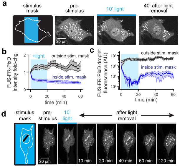
Figure 3. PixELLs exhibit long-term spatial memory of transient stimuli.
(a) Schematic and images of spatially-restricted 450 nm light stimulation. Fluorescent images of FUSN-FusionRed-PixD are shown for cells before, during and after stimulation. (b) Cytoplasmic intensity in regions inside and outside the stimulation mask for 4 cells. Mean ± SEM are shown. (c) Mean cluster size for the cell in a, averaged across 5 clusters inside and outside the stimulation area. (d) Still images showing long-term memory of a nucleus-localized light stimulus. See also Figure S4 and Movie S6.

