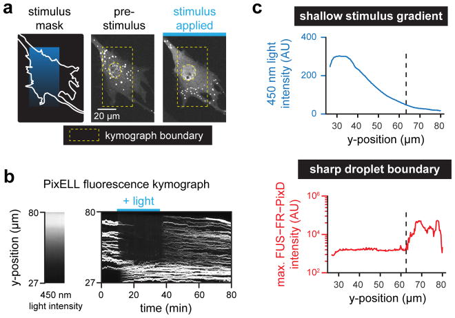
Figure 4. PixELLs amplify shallow stimulus gradients into all-or-none spatial patterns of droplets.
(a) Gradient stimulation of a PixELL-expressing NIH3T3 cell. Fluorescent images of FUSN-FusionRed-PixD are shown for a representative cell stimulated with a linear gradient of light intensity. (b) Kymograph of maximum FUSN-FusionRed-PixD fluorescence within each row of the yellow box from a (right), and median blue light intensity measured within the yellow box from a (left). (c) Quantification of the kymograph in b at 35 min, after spatial light pattern is established. A gradual decrease in 450 nm intensity (top panel; blue curve) elicits a sharp, switch-like transition to form bright FUSN-FR-PixD droplets (bottom panel; red curve). See also Movie S7.

