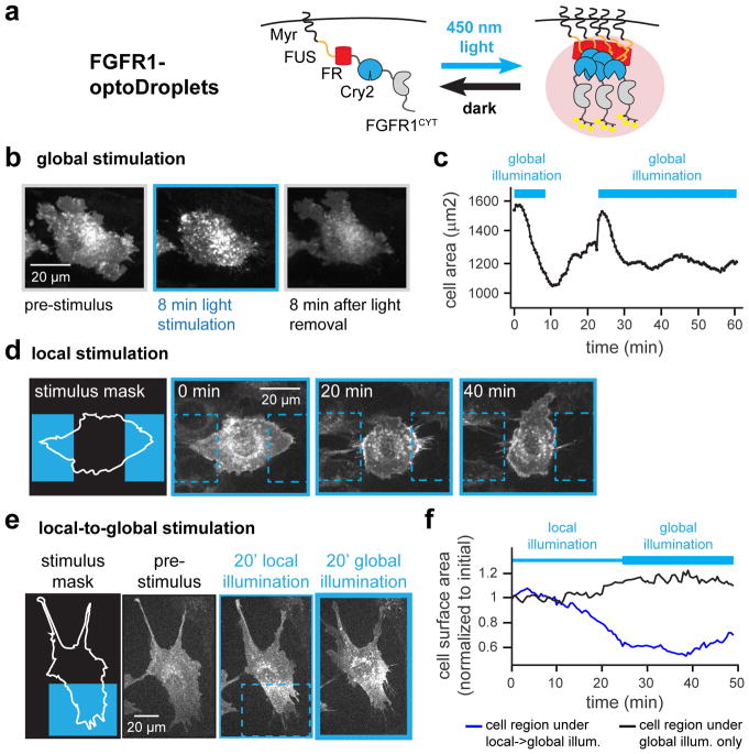
Figure 6. Liquid phase separation drives spatial memory in RTK signaling.
(a) Schematic showing FGFR1-optoDroplets for inducing RTK clustering and downstream signaling. (b) FGFR1-optoDroplet cells reversibly “cringe” in response to global blue light stimulation. (c) Quantification of change in cell surface area for cell pictured in b. (d) FGFR1-optoDroplet cells retract in response to light, ‘avoiding’ a local light stimulus. (e) FGFR1-optoDroplet cells exhibit persistent local clustering and cytoskeletal contraction even after a switch to global illumination. (f) Quantification of cell surface area within the local-to-global illuminated region (blue box in e) and global-only illuminated region (remainder of cell in e) during local-to-global illumination. See also Figure S5 and Movies S10–S12.

