
Figure 3.
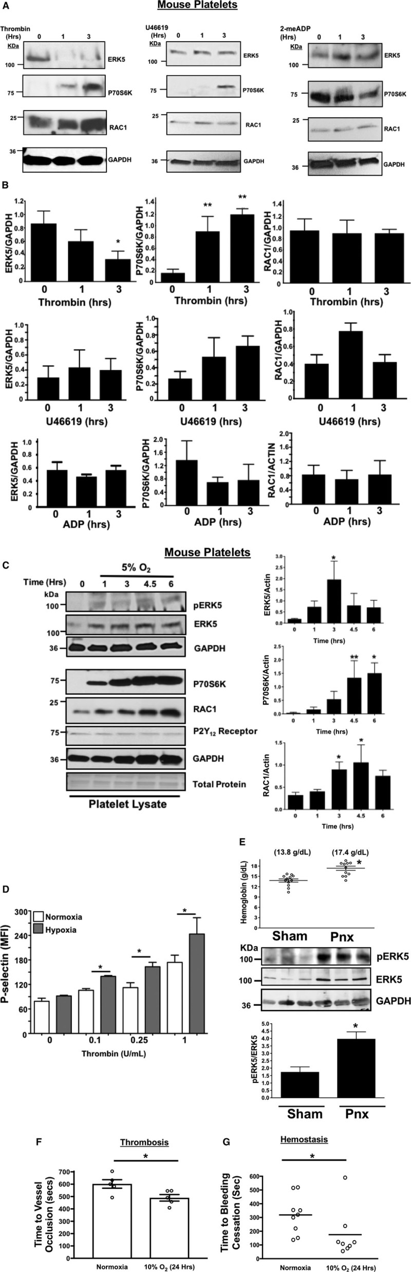
Murine platelet protein expression changes in vitro after agonist stimulation. A and B, Platelets isolated from mice were stimulated with 0.2 U/mL thrombin, 10 µmol/L U46619, or 10 µmol/L 2-me-ADP for 1 to 3 h. Protein expression was determined by (A) immunoblot (IB) and (B) quantified by densitometry (mean±SEM, *P=0.12 vs 0 for ERK5 (thrombin) and **P<0.05 vs 0 for P70S6K (thrombin) by 1-way ANOVA, n=3). Platelets isolated from mice were incubated for 0 to 6 h under hypoxic conditions (5% O2) and platelet protein expression was assessed by IB and quantified by densitometry (mean±SEM, *P<0.05 or **P=0.058 vs 0 by 1-way ANOVA, n=4–6). D, Platelets were isolated from wild-type (WT) mice and activation was assessed after 6 h of normoxia or hypoxia and stimulated with thrombin for 15 min. Activation was assessed by P-selectin expression (mean±SEM, n=4, *P<0.05 by 1-way ANOVA). E, Left pneumonectomy (Pnx) or sham surgery mice demonstrate hypoxia by a compensatory increase in circulating blood hemoglobin concentration, P=0.0002 by t test between groups without a change in platelet count (559±44 vs 604±43 between groups, P=0.48 by t test, n=12 in each group). Isolated platelets show platelet ERK5 (extracellular regulated protein kinase 5) activation (mean pERK5/ERK5±SEM, *P=0.009 by t test, n=4 in each group). Mesenteric artery thrombosis was assessed in mice after living for 24 h at ambient oxygen or in hypoxic conditions (10% O2, mean time to vessel occlusion±SEM, n=5 in each group, *P=0.026 by 1-way ANOVA for normoxia vs 10% O2). G, Tail bleeding times as an index of hemostasis were calculated as the time in seconds for bleeding to stop after surgical amputation of the tail tip, median value, n=8 to 9, *P=0.021 by Mann–Whitney U test for normoxia vs 10% O2.