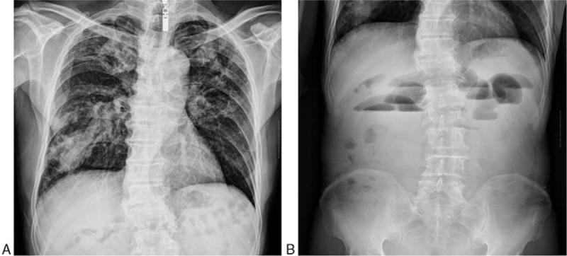
Figure 1.

Plain x-rays of chest and abdomen on hospital admission. (A) Chest x-ray showing bilaterally patchy infiltrates, increased bronchovascular markings, and mass in the right lower lung fields. (B) Abdomen x-ray showing showed the gaseous distention of the bowel with air-fluid levels.
