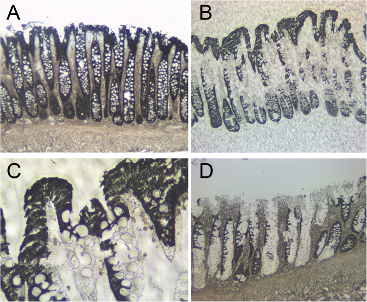
Figure 1.
Isolation of colonic epithelial cells using xMD. (A) A slide undergoes IHC for the cytokeratin AE1/AE3, a common antibody for epithelial cells. This slide is not coverslipped or counterstained. (B) After xMD, the pigmented cells are selectively transferred onto an EVA membrane. (C) High-power view of the EVA membrane demonstrating the tight isolation of the pigmented epithelial cells. (D) The original slide, after xMD, shows an incomplete transfer of epithelial cells, but no transfer of lamina propria or stromal areas (Images in A, B & D are 100x original magnification while image C is 400x original magnification).

