
Figure 4.
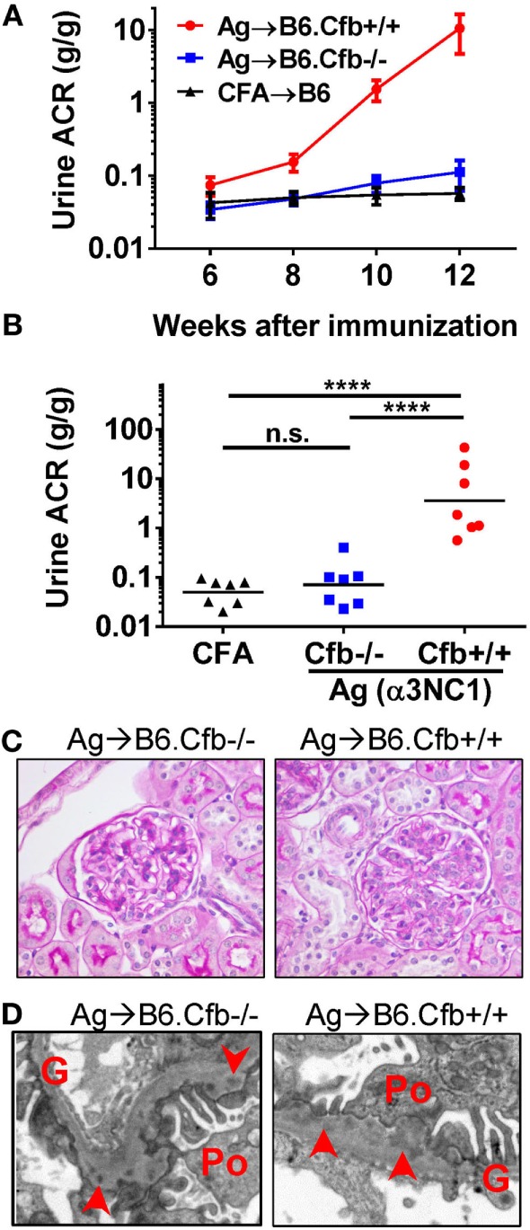
B6.Cfb−/− mice immunized with α3NC1 are protected against albuminuria despite developing subepithelial immune complexes. (A) Time course of urinary albumin-to-creatinine ratio (ACR) in B6.Cfb+/+ mice (circles) and B6.Cfb−/− mice (squares) immunized with α3NC1 (N = 7 per group). Control B6 mice (triangles), including both genotypes, were immunized with adjuvant alone (N = 7). Shown are means and SEM. (B) Scatterplot depicts the ACR values at the endpoint of this experiment (week 12). The significance of differences among groups was analyzed by one-way ANOVA with Dunnett’s correction for multiple comparisons. ***P < 0.001, ****P < 0.0001. n.s., not significant. (C) Morphology of kidneys from α3NC1-immunized B6.Cfb−/− and B6.Cfb+/+ mice appears normal by light microscopy (periodic acid–Schiff staining, original magnification 400×). (D) Transmission electron microscopy shows subepithelial electron dense deposits (arrowhead), expansion of the glomerular basement membrane (G), and podocyte (Po) foot process effacement. Original magnification 7,500×.