
Figure 3.
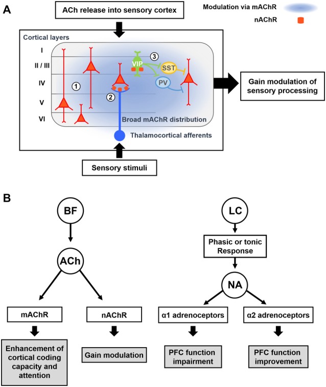
Modulatory effects of acetylcholine (ACh) and Noradrenaline (NA) on cortical processing through different types of receptors. (A) Schematic description of cholinergic modulation in the primary sensory cortex. ① Broad distribution of metabotropic ACh receptor (mAChR) mediates modulation of both excitatory and inhibitory neurons across the layers (Alitto and Dan, 2013). ② nicotinic AChRs (nAChRs) are expressed in the thalamocortical axon terminals, and cholinergic activation of them causes increase in sensory responses of neurons in the input layer (Lavine et al., 1997; Metherate, 2004; Disney et al., 2007). ③ nAChRs are expressed in the vasoactive intestinal peptide-positive (VIP+) neurons, which elicit disinhibition of pyramidal neurons by inhibiting SST+ or PV+ inhibitory neurons (Harris and Mrsic-Flogel, 2013; Lee et al., 2013; Pi et al., 2013). Cholinergic activation of VIP+ neurons can increase the sensory gain via this disinhibitory circuit (Porter et al., 1999; Fu et al., 2014). (B) Modulatory effects of ACh and NA. (Left) ACh released from the BF enhances cortical processing via both mAChRs and nAChRs. Activation of mAChR enhances cortical coding capacity of sensory stimulus (Goard and Dan, 2009), while activation of nAChRs increases the sensory gain in the visual cortex (Metherate, 2004; Disney et al., 2007). (Right) Two distinct modes of the noradrenergic modulation. LC neurons show either phasic or tonic activity patterns depending on the states. Sensory stimuli evoke phasic responses of NA neurons whereas stressful stimuli evoke both phasic and tonic responses (Aston-Jones et al., 1999). When the animal shows focused attention or engages in the task, the NA neurons show phasic activity. Conversely, NA neurons show tonic responses when the animal is distracted or shows flexible behaviors. The amount of NA released from noradrenergic neurons determines the activation of different types of adrenoceptors, which modulate the PFC function in an opposite manner (Ramos and Arnsten, 2007). A moderate amount of the NA preferentially activates the α2 adrenoceptor, which has a higher binding affinity for the NA, and improves the PFC function such as working memory and focused attention (Li and Mei, 1994; Li et al., 1999). In contrast, when a higher concentration of NA is released, the α1 adrenoceptors are activated as well, which can lead to the impairment of PFC function (Arnsten et al., 1999; Mao et al., 1999; Ramos and Arnsten, 2007).