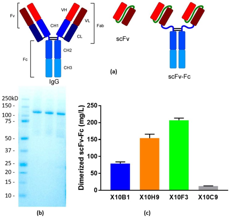
Figure 3.
Cell-free expression of candidate scFv-Fc antibody fragments. (a) Representation of the structure of the three antibody fragments utilized in this study. The typical IgG format is shown on the left, the scFv format utilized for binding and neutralization in the center, and the scFv-Fc format utilized in neutralization and protection studies on the right. (b) Coomassie stained SDS-PAGE Gel of purified scFv-Fcs. Precision Plus Protein™ Standards (BioRad, Hercules, CA, USA); Lane 2: X10F3; Lane 3: X10H2; Lane 4: X10B1. (c) Titers from the cell-free expression of the candidate scFv-Fcs.

