Figure 2.

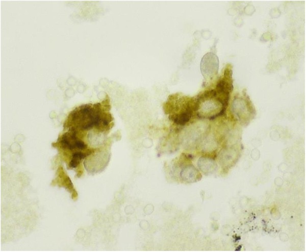
Immunohistochemical stain, performed on the cellblock, shows that the neoplastic cells are positive for calcitonin (cytoplasmic and granular staining; high power ×60 magnification).

Immunohistochemical stain, performed on the cellblock, shows that the neoplastic cells are positive for calcitonin (cytoplasmic and granular staining; high power ×60 magnification).