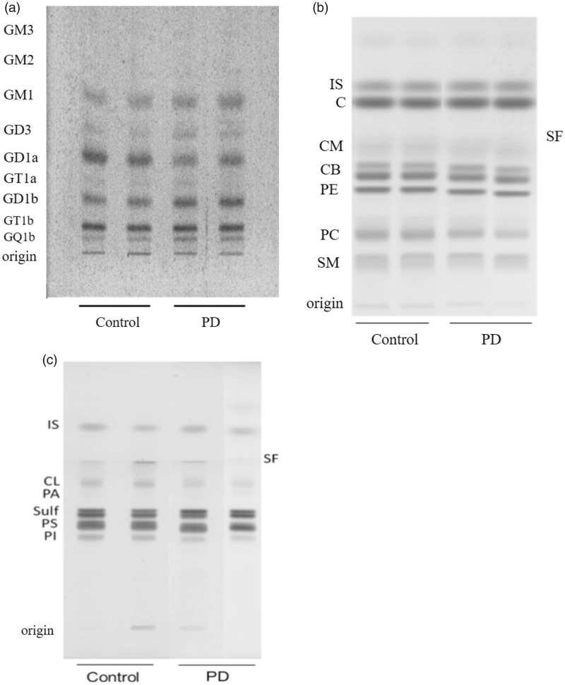
Figure 1.
(a) HPTLC of gangliosides in substantia nigra (SN) samples from male control and PD subjects. The amount of ganglioside sialic acid spotted per lane was equivalent to approximately 1.5 µg. The plate was developed by a single ascending run with CHCl3:CH3OH:dH2O (55:45:10 by vol) containing 0.02% CaCl2·2H2O. The bands were visualized with the resorcinol-HCl spray, as described previously (Hauser et al., 2004). HPTLC of SN neutral lipids (b) and acidic lipids (c) in male control and PD subjects. The amount of neutral lipids and acidic lipids spotted per lane was equivalent to approximately 35 µg and 100 µg tissue dry weight, respectively. The plates were developed as we described previously (Baek et al., 2009). The second PD sample in (c) was moved to its position from another region on the same HPTLC, which explains the merge line seen on the plate. Neutral lipids include: CE = cholesteryl esters; TG = triglycerides; IS = internal standard; C = cholesterol; Cer = ceramide; CB = cerebrosides (doublet); PE = phosphatidylethanolamine; PC = phosphatidylcholine; SM = sphingomyelin. As the zwitterionic lipids (PC, PE, and SM) elute with the neutral lipids, they are included in this group. Acidic lipids include: FA = fatty acids; IS = internal standard; CL = cardiolipin; PA = phosphatidic acid; SULF = sulfatides (doublet); PS = phosphatidylserine; PI = phosphatidylinositol.

