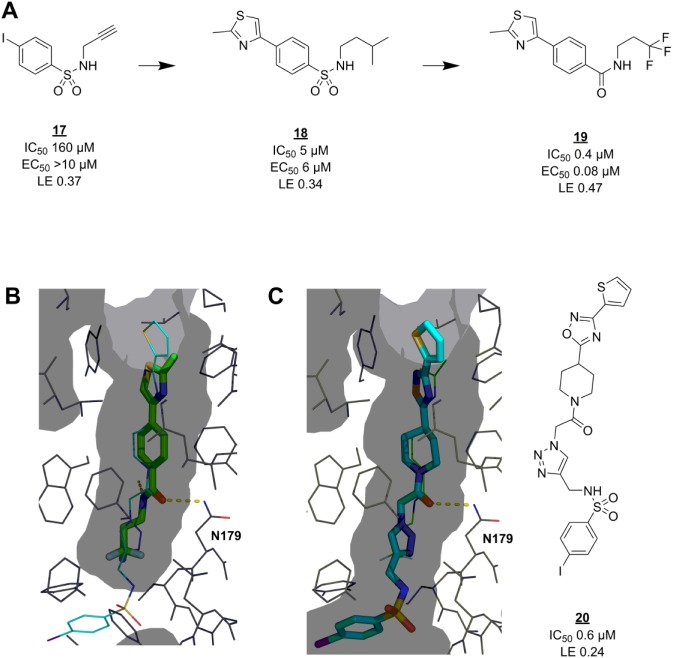
Figure 4.
Fragmentation approach for EthR inhibitors. (A) Click reaction component 17 can be seen as a fragment with weak activity. It was successfully grown via virtual (18) and then actual medicinal chemistry to lead compound 19. (B) Crystal structure (PDB code 4M3B86) showing the binding mode of 19 (green sticks) in the M. tuberculosis EthR allosteric pocket (grey surface representation). The binding pose of 20 (cyan thin lines) in the same pocket (PDB code 3O8H85) is overlaid. (C) Crystal structure (PDB code 3O8H) showing the binding mode of 20 (cyan sticks) in its binding pocket (grey surface). The binding pose of 19 (green thin lines) in the same pocket (PDB code 4M3B) is overlaid. Hydrogen bonds are shown as dashed yellow lines. EC50 = concentration of EthR ligand at which M. tuberculosis growth in macrophages is inhibited by 50% by ethionamide at 1/10 of its MIC, determined according to a standard procedure.106

