
Figure 4.
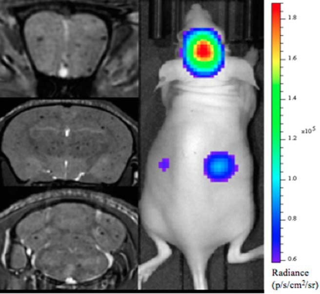
Bioluminescence (BLI) and MRI of a representative mouse from group 3 on day 0. Whole mouse BLI shows strong signal in the brain and weaker signal in the mouse body (Right Panel). MR image slices from a 3-dimensional (3D) bSSFP image acquisition for the same mouse (Left Panel). The 3 images are at the level of the frontal brain, midbrain, and hindbrain, and signal voids are detected throughout.