
Figure 5.
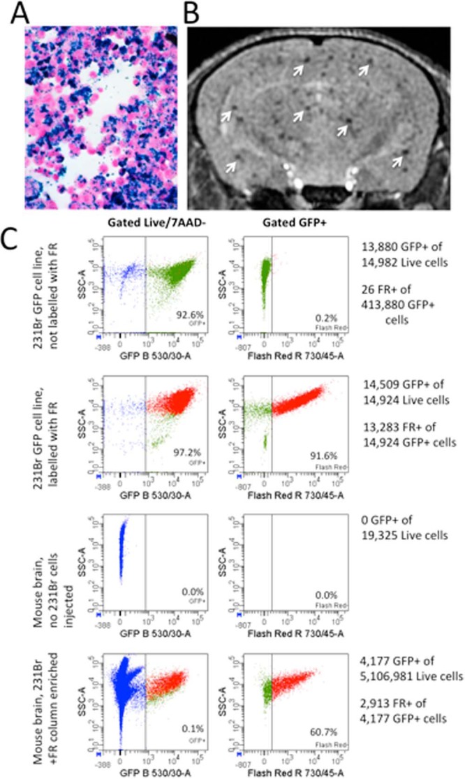
Isolation of live green fluorescent protein (GFP)+ Flash Red+ cells from whole mouse brain. Perl's Prussian Blue staining (blue) confirmed MPIO labeling of 231BR cells; 10× magnification (A). MRI confirmed the successful delivery of MPIO-labeled 231BR cells to mouse brain (arrows) (B). FACS analysis of dissociated mouse brain concentrated with a magnetic column identified and isolated a subset of live GFP+ Flash Red+ cells from the dissociated brain cell population. GFP+ Flash Red+ cells were only found and isolated from mice that had received labeled 231BR cell injections (C).