Figure 2.

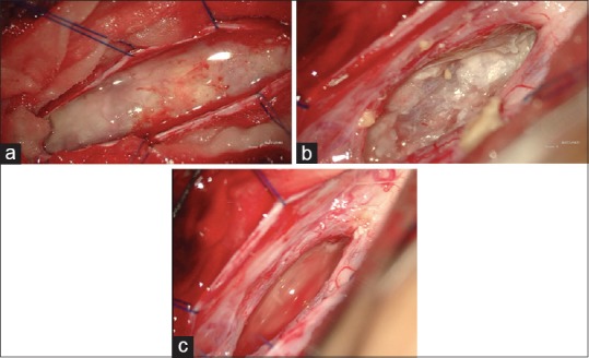
Intraopeartive images. (a) Lesion surfacing after durotomy, (b) pearly white material seen during decompression of the tumor and (c) tumor cavity after decompression

Intraopeartive images. (a) Lesion surfacing after durotomy, (b) pearly white material seen during decompression of the tumor and (c) tumor cavity after decompression