Figure 3.

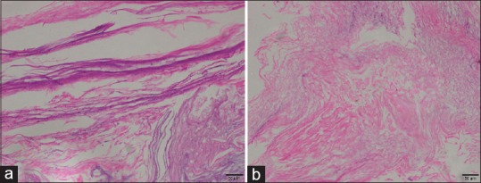
Histological images. (a and b): Photomicrograph of an epidermoid cyst with lamellated keratin. Adnexal structures were not seen. (H&E, ×100)

Histological images. (a and b): Photomicrograph of an epidermoid cyst with lamellated keratin. Adnexal structures were not seen. (H&E, ×100)