Figure 2.
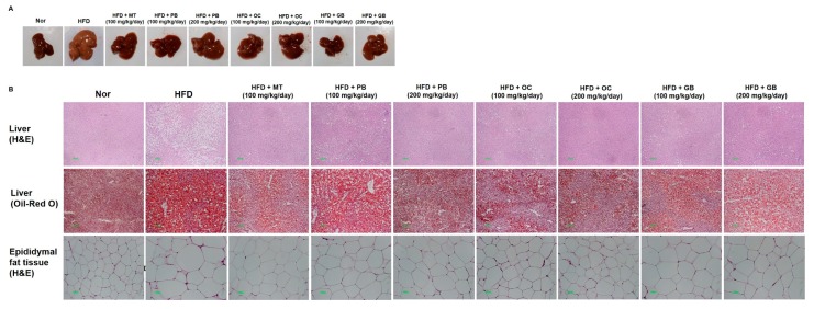
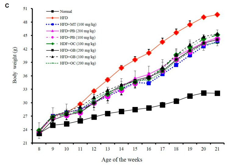
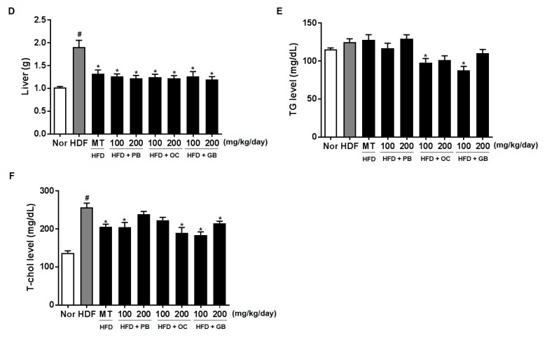
Effects of PB, OC, and GB extracts on mouse liver histology and body weight in a high-fat diet (HFD)-fed mouse model. (A) Photographs of the mouse liver. (B) Histopathological examination by hematoxylin and eosin (H&E) (liver and epididymis fat tissue) and Oil Red O (liver) staining and (C) body weights are shown for HFD-fed mice, with or without PB, OC, and GB extract supplementation. Liver weights (D), TG levels (E), and total cholesterol (TC) levels (F) in the liver tissues from the mice treated with the PB, OC, and GB extracts. MT, milk thistle. The data are presented as the mean ± SEM (n = 9); # p < 0.05 compared with the control group; * p < 0.05 compared with the HFD group.


