Figure 1.
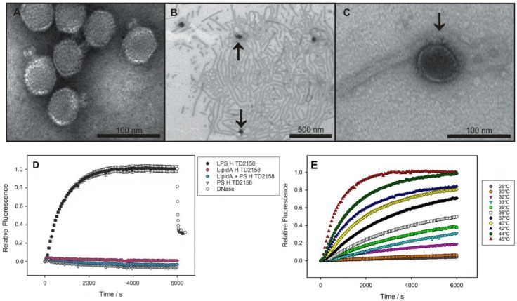
LPS-mediated DNA-release from phage HK620 observed in vitro. (A) Transmission electron microscopy images of phage HK620 particles (1 × 10¹⁰ pfu mL−1) stained with 1% (w/v) uranyl acetate and pre-incubated with 0.24 mg mL−1 LPS from E. coli H TD2158 (O18A1) at 37 °C (B,C). LPS from E. coli H TD2158 (O18A1) forms ribbon-like aggregates (B) and phages are attached to these LPS structures (arrows). (D) DNA ejection from phage HK620 (6.7 × 10⁹ pfu) triggered by 16.7 μg/mL E. coli H TD2158 (O18A1) LPS in the presence of the fluorescent DNA-binding dye Yo-Pro at 45 °C (black circles). Ejected DNA could be digested by adding DNase after 6000 s (white circles). Controls contained an E. coli H TD2158 (O18A1) lipid A preparation (pink circles), an O-antigen polysaccharide preparation lacking lipid A (grey triangles) or a mixture of both (blue circles). Standard deviations were obtained from three independent experiments and do not exceed 5% of the total fluorescence signal. (E) DNA ejection of HK620 at different temperatures (same conditions as in D).

