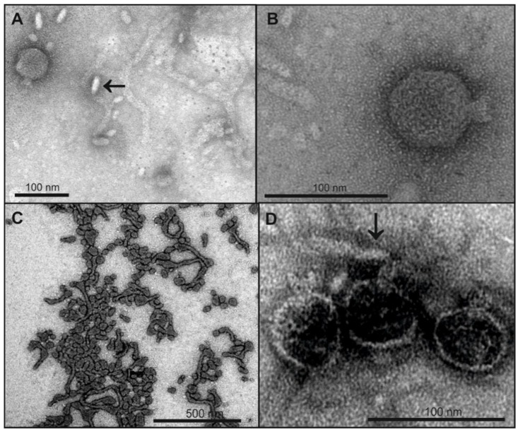
Figure 4.
Legend: TEM micrographs of phage HK620 before and after incubation with non-host E. coli LPS. 1 × 10¹⁰ pfu mL−1 HK620 particles were stained with 1% (w/v) uranyl acetate after overnight incubation at 37 °C with 0.24 mg mL−1 LPS from E. coli IHE3042 (O18A1) (A,B) or E. coli DSM 10809 (O18A) (C,D). For IHE3042 (O18A1) with short O-antigen chains mainly small LPS structures are visible (arrow in A) and HK620 particles are not attached to these structures (B), whereas on DSM 10809 (O18A) LPS with longer O-antigen chains empty HK620 particles are attached (D, arrow).

